We work at the interface of bioanalytical chemistry and aerosol science.
Our lab builds instrumentation and develops quantitative analytical methods to study disease-related biochemistry and develop new approaches to diagnosis and treatment. A major focus of our research is the respiratory system, including noninvasive sampling and aerosol-based technologies.
Our Research
Noninvasive Sampling of the Deep Lung
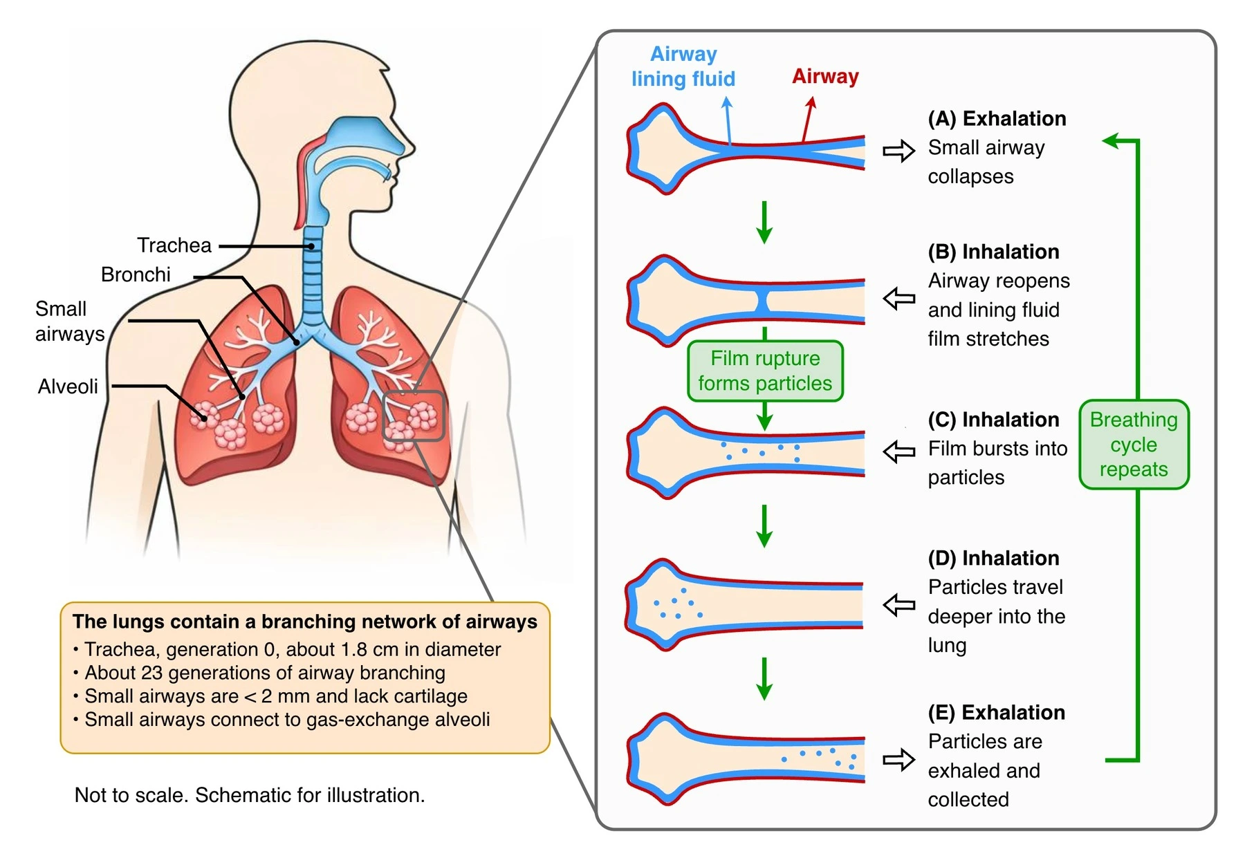
Text description of the airway diagram
The schematic labels the trachea, bronchi, small airways and alveoli. The trachea (generation 0) is about 1.8 cm in diameter. The lungs have about 23 generations of airway branching. Small airways are less than 2 mm across, lack cartilage, and connect to gas-exchange alveoli. The drawing is not to scale.
- Exhalation: a small airway collapses.
- Inhalation: the airway reopens and the airway lining fluid stretches into a film.
- Continued inhalation: the film ruptures, forming fluid particles.
- Particles travel deeper into the lung during inhalation.
- On exhalation, particles leave the lung and can be collected. The breathing cycle repeats.
Arrows show the sequence. Blue identifies airway lining fluid and particles; red outlines the airway. The labels and sequence carry the same information without relying on color.
Every breath repeatedly opens and closes the smallest airways deep in the lung, rupturing a thin fluid film into micron-scale droplets. We build the instrumentation and analytical methods to capture and quantify these particles for disease diagnosis and monitoring.
See our research →Recent Publications
We Are Building a Team
We are actively recruiting graduate students and undergraduate researchers eager to work at the intersection of analytical chemistry, biochemistry, and device engineering. If you are excited about building new tools to detect disease, we want to hear from you.
Open Positions →